Skin DiseasesDermatofibroma: Causes, Symptoms, Diagnosis, and Treatment

Dermatofibroma: Causes, Symptoms, Diagnosis, and Treatment in Estonia

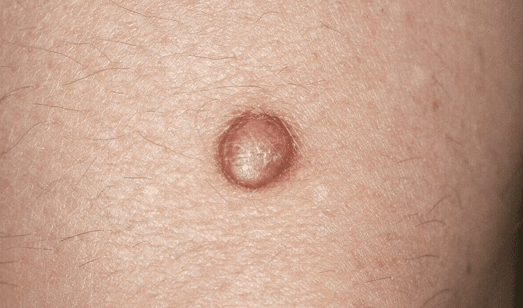
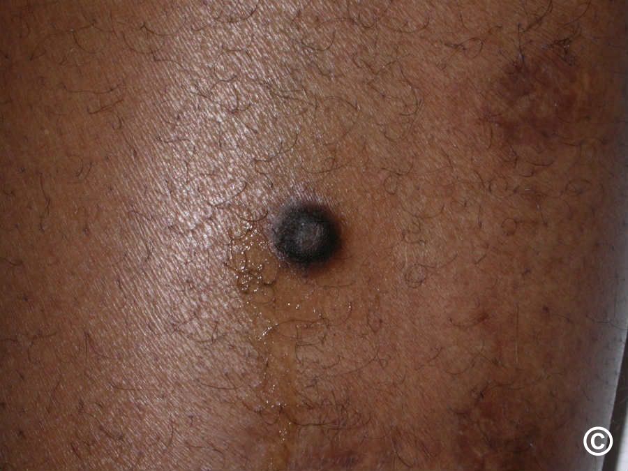
A small dome-shaped brownish dermatofibroma on the skin – a firm, benign nodule.

Need an online dermatologist for dermatofibroma in Estonia? iDerma's doctors review your photos and reply within 24 hours — from 49 €.

Dermatofibroma (also called histiocytoma) is a common benign skin growth arising from a localised proliferation of fibroblasts and histiocytes in the dermis. It develops most often on the lower limbs, occurs across all skin tones, and is more frequent in women — typically presenting in the 20–50 age range.

In many cases, dermatofibromas develop after minor skin trauma — an insect bite, needle prick, shaving cut, or thorn scratch. They are also more common in people with a weakened immune system, such as those living with HIV or autoimmune conditions.
Clinical Features

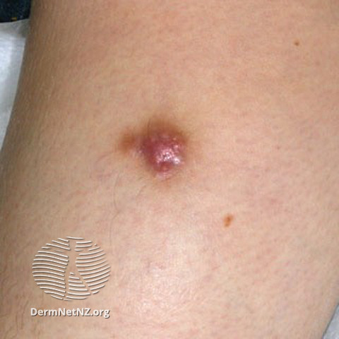
Dermatofibromas typically present as a firm, slightly raised or flat nodule with a smooth surface. Colour varies from pink or light brown on fair skin to dark brown or black on darker skin tones; a lighter area is sometimes visible at the centre. Size ranges from 0.5 to 1.5 cm, most commonly 7–10 mm.

Most lesions cause no symptoms. Some people, however, notice:

  • A tightness or pulling sensation over the growth
  • Pain or tenderness to touch
  • Itching
  • Increased local sensitivity

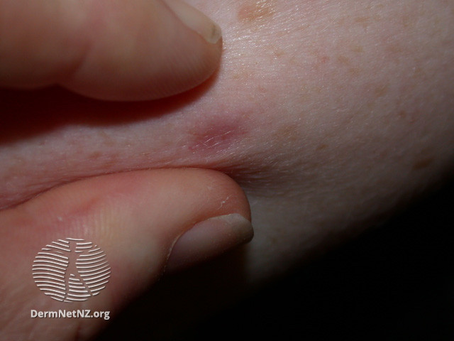
A reliable bedside finding is the dimple sign (pinch sign): gentle squeezing of the skin on both sides of the lesion causes the centre to dimple inward. This happens because the growth is tethered to the deeper layers of the skin.

Diagnosis

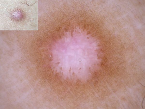
Dermatofibroma is usually diagnosed through clinical examination alone. Dermoscopy — a non-invasive magnified skin examination — helps a dermatologist assess the lesion's internal structure and distinguish it from other growths.

In atypical or uncertain cases, surgical excision with histological analysis (skin biopsy) provides a definitive diagnosis and removes the lesion at the same time.
Treatment

Because dermatofibromas are harmless, treatment is not always needed. Removal is considered when the growth causes persistent discomfort, when histological confirmation is required, or when the patient requests it for cosmetic reasons.

Available methods:

  • Surgical excision — the most thorough approach; recurrence is uncommon but a visible scar usually remains
  • Cryotherapy — liquid nitrogen freezing; lower scar risk, but higher chance of recurrence
  • Electrosurgery — useful for superficial lesions
  • Laser therapy — may reduce pigmentation but rarely removes the full depth of the growth

When to See a Dermatologist

Dermatofibroma can look similar to certain malignant skin tumours, particularly melanoma. Any skin growth that is new, enlarging, changing colour, or symptomatic warrants professional assessment. A dermatologist will use dermoscopy to evaluate the lesion and, if there is any doubt, arrange a skin biopsy to rule out malignancy definitively. Book an online dermatologist consultation if you have a skin growth you would like assessed.


Frequently asked questions

Is dermatofibroma dangerous or cancerous?
Dermatofibroma is benign and not cancerous. It poses no direct health risk. The main concern is its visual resemblance to certain malignant tumours — particularly melanoma. Any skin growth that changes in size, colour, or shape should be assessed by a dermatologist rather than watched at home.
Can dermatofibroma go away on its own?
Most dermatofibromas persist for many years without resolving. Spontaneous regression is occasionally reported but uncommon. If the lesion is stable, confirmed benign, and causing no symptoms, watchful waiting is a reasonable approach — but a confirmed diagnosis comes first.
What is the dimple sign in dermatofibroma?
The dimple sign (pinch sign) is the hallmark of dermatofibroma: when you gently pinch the skin on either side of the lesion, the centre dimples inward instead of protruding. This happens because the growth is attached to the deeper layers of the skin. The sign is reliable but a dermatologist will use dermoscopy for a full assessment.
Will there be a scar after dermatofibroma removal?
Surgical excision, the most complete method, typically leaves a visible linear scar that may not fully meet cosmetic expectations. Cryotherapy and laser therapy tend to produce less scarring but carry a higher recurrence rate. Your dermatologist can help weigh the options based on the lesion's size, location, and your priorities.
How do I know if my skin growth is a dermatofibroma?
Only a dermatologist can confirm the diagnosis reliably. The dimple sign, typical size (0.5–1.5 cm), and a stable history are reassuring features. Dermoscopy allows a detailed look at the lesion's internal pattern. If any doubt remains, a small skin biopsy provides a definitive answer. Do not rely on self-assessment for a firm skin nodule — get it checked.
Still unsure?

A dermatologist writes a plan made for your skin.

Not another off-the-shelf cream — a certified specialist’s diagnosis and personal treatment plan, within 24 hours.

Start your consultation
Personal treatment plan
24h
Diagnosis
Treatment plan
Prescriptions
iDerma
Board-certified dermatologist

Article by

Anna Tunkeviča

Medical content reviewed by

Andra Šukienė(Dermatologist)

Our other articles

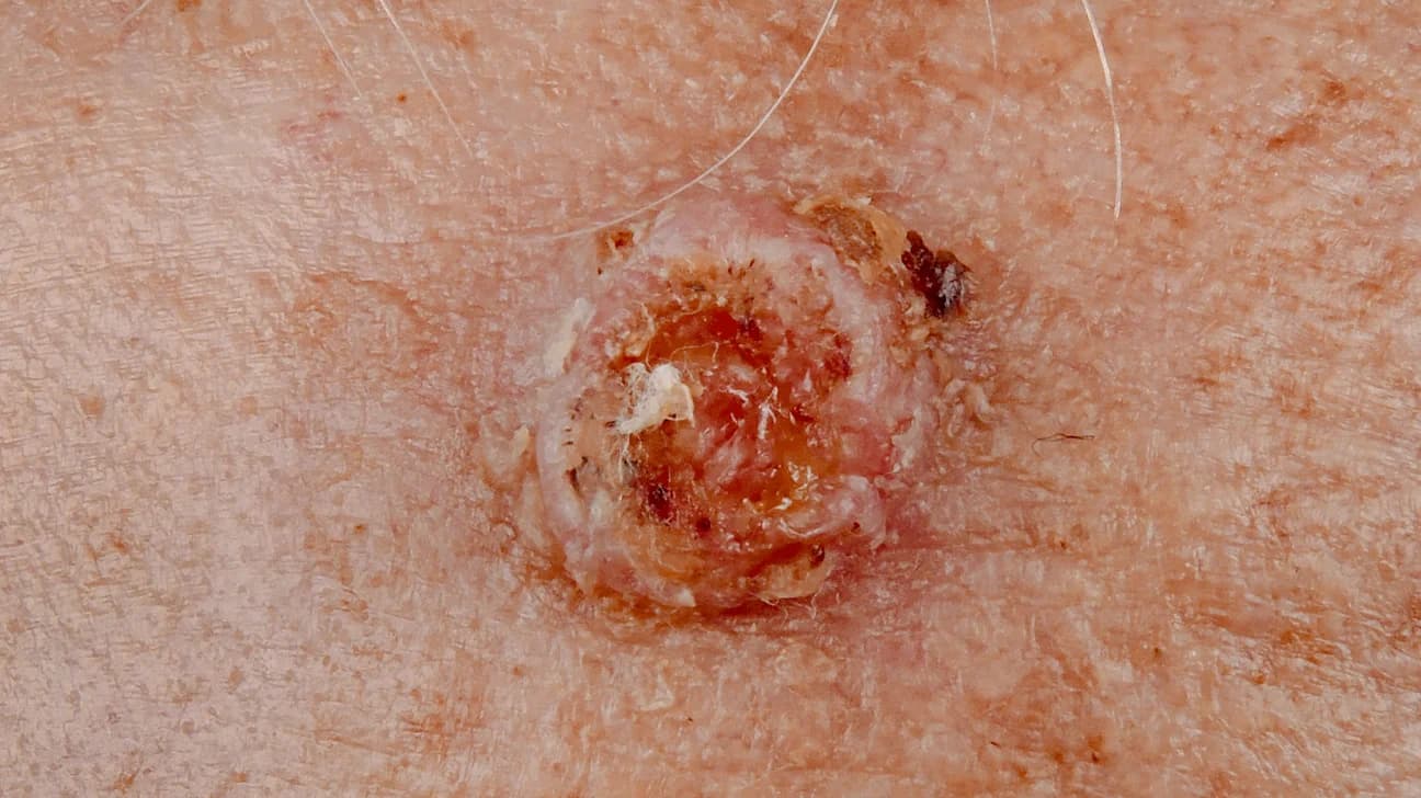
Crusted, ulcerated nodule on the skin, characteristic of squamous cell carcinoma, a form of skin cancer.

Squamous cell carcinoma: causes, symptoms, diagnosis, and treatment

Learn what squamous cell carcinoma is, how to recognize it, when to see a dermatologist, and what treatment and prevention options are available.
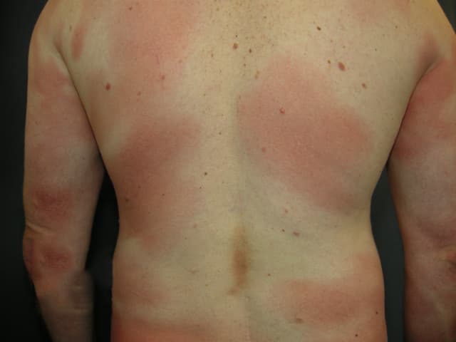
Large reddish patches across the back and torso typical of the patch stage of mycosis fungoides

Mycosis fungoides: symptoms, stages & treatment options

Mycosis Fungoides is a slow-growing T-cell skin lymphoma causing red scaly patches. Early diagnosis and care help manage symptoms effectively.
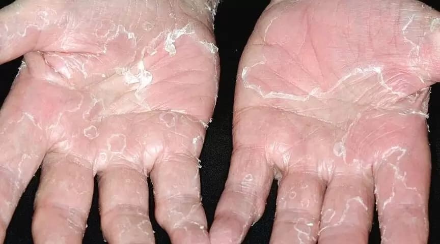
Both palms with peeling skin syndrome, skin shedding in large sheets to reveal reddened skin underneath.

Peeling skin syndrome: symptoms, causes, treatment

Rare genetic disorder causing continuous skin peeling. Learn symptoms, diagnosis & treatment. Manageable with proper care. Guide for patients & families.
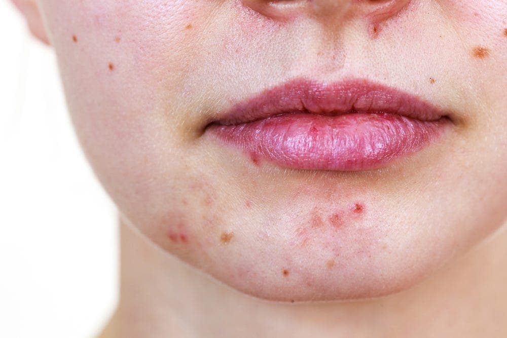
Adult woman's acne with breakouts and post-inflammatory marks around the lips and on the chin

Adult acne: causes, symptoms, and effective treatment

Discover what causes adult acne, how to recognize the symptoms, and which treatments and skincare tips can help you manage breakouts effectively.